HIGHLIGHTS
Montefiore Einstein One of Two North American Institutions to Use Intraoperative Cutting-Edge PET-CT Imaging in Head and Neck Cancer Surgery

Montefiore Einstein is one of just two institutions in North America enrolling patients in a clinical trial to demonstrate the effectiveness of the Aura 10 PET-CT imager, a new intraoperative imaging technology. This groundbreaking technology enables surgeons to examine resected head and neck cancer tissue specimens in real time in the operating room, aiming to provide a more comprehensive, three-dimensional view of tissue margins to improve surgical accuracy and the chance of complete resection.
“With this imaging technology, we are aiming to optimize cancer outcomes and functional outcomes for patients—taking that ‘Goldilocks’ amount of tissue: not too much and not too little,” said Vikas Mehta, MD, MPH, FACS, Vice Chair, Otorhinolaryngology – Head and Neck Surgery, Montefiore Einstein and Associate Professor, Otorhinolaryngology – Head and Neck Surgery, Albert Einstein College of Medicine. Dr. Mehta is lead investigator on the active clinical trial that brought the Aura 10 to Montefiore Einstein.

Vikas Mehta, MD, MPH, FACS
Tumor margins are crucial to cancer outcomes
Successful resective surgery includes removal of cancerous tissue plus surrounding healthy tissue to create negative (cancer-free) margins. Surgeons must visualize estimates on the tumor’s extension based on preoperative imaging and plan surgery accordingly. Despite cancer surgeons’ best efforts to perform complete resections, 20% to 30% of solid-tumor resections in various types of cancer do not achieve negative margins.
The final determination of margin status is made by microscopic analysis of histopathology, which takes place in the lab postoperatively[GC1] ; results can take up to a week. If the excised tumor’s margins are determined to be positive, patients typically undergo intensive radiotherapy and/or chemotherapy, but these treatments only partly mitigate the increased risk of cancer recurrence.
“The margins really matter for the patient. If we leave positive margins, the chance of cancer recurrence and of the patient dying from the tumor is double or triple the amount it is if the tumor gets cleared,” said Dr. Mehta. “We hope that this imaging will improve our ability to assess and address tumor margins in real time.”
The intraoperative process
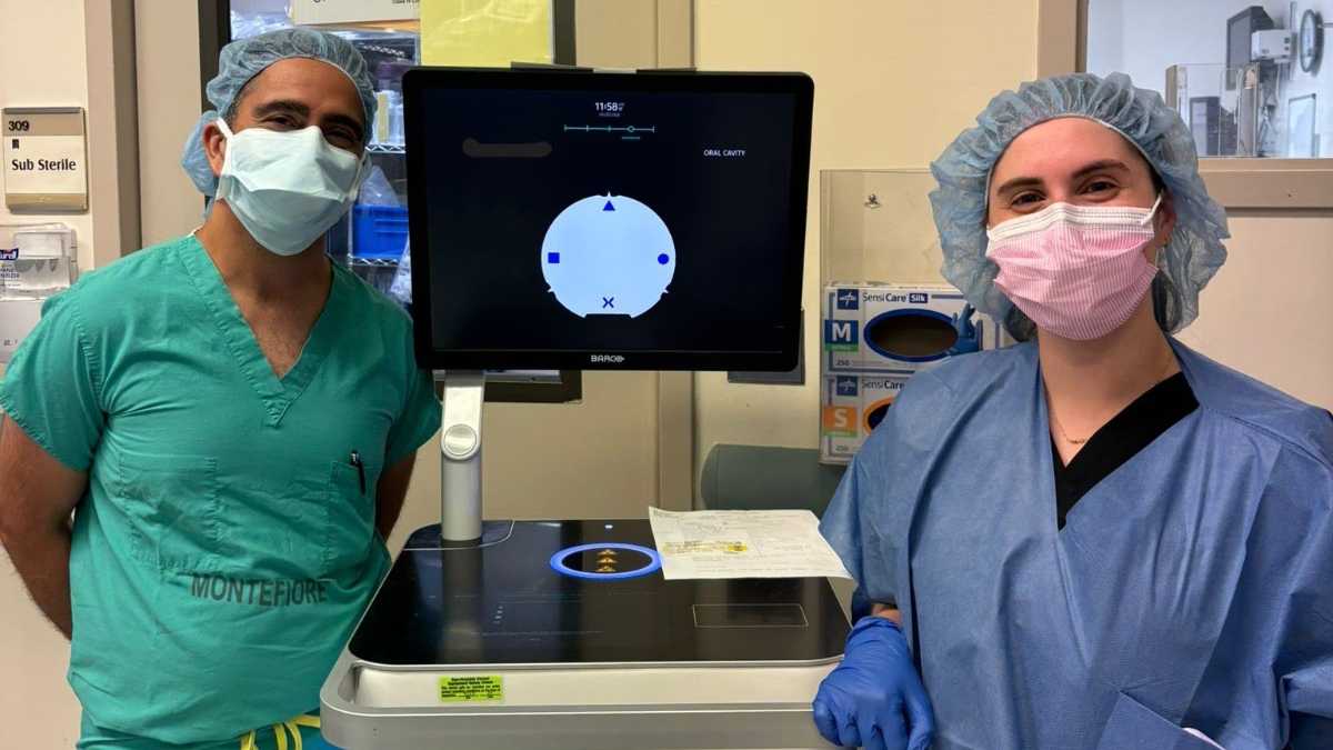
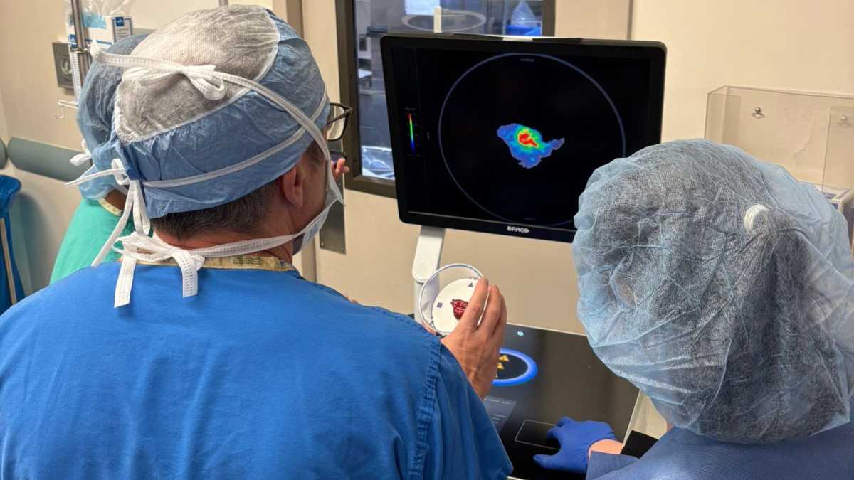
The Aura 10 uses the same positron emission tomography (PET) and computed tomography (CT) imaging technologies that are used in PET-CT scans performed in more traditional settings. But instead of scanning the patient’s body in a PET-CT machine, only the resected tumor is scanned in the mobile imaging unit—all while in the operating room.
Patients who opt for this imaging during their surgery receive an injection of a glucose solution with a radioactive tracer, which is absorbed more readily by cancer cells than by surrounding tissues. Tumor resection proceeds as usual, and then the resected tumor is placed in a special cassette and scanned by the Aura 10. The mobile PET-CT specimen imager analyzes the sample, which takes approximately 10 minutes, and areas with cancer cells present are highlighted on the resulting high-resolution images, including an interactive 3D [GC2] image that can be rotated on demand and viewed from any angle.
Reviewing the images in real time gives surgeons additional feedback about the margin status while validating the surgeon’s confidence in a complete resection or providing the opportunity to make an informed decision about how to proceed.
A promising clinical trial
The Aura 10 was cleared by the Food and Drug Administration in 2023, and a clinical trial is now under way to investigate its feasibility and compare it to the gold standard [GC3] of histopathology specimen analysis. Dr. Mehta and others at Montefiore Einstein are collaborating with a team at Vanderbilt University in Nashville, Tennessee, which is the only other institution in North America where this technology is available to patients.
The trial aims to enroll at least 20 participants at Montefiore Einstein over an 18-month period. Six patients have undergone surgery that utilized intraoperative PET-CT, with two more cases planned in the future.
Dr. Mehta is working closely with Bradley A. Schiff, MD, Director, Head and Neck Surgery, Otorhinolaryngology – Head and Neck Surgery, Montefiore Einstein and Professor, Otorhinolaryngology – Head and Neck Surgery, Albert Einstein College of Medicine, who is also enrolling patients in the study and performing surgical cases that are part of the clinical trial. Renee M. Moadel, MD, MSc, Attending Physician, Radiology, Montefiore Einstein and Assistant Professor, Nuclear Medicine and Medicine, Albert Einstein College of Medicine; research nurse Stelby Augustine, RN; and medical student Sara Friedman, Albert Einstein College of Medicine Class of 2027, have all been instrumental in the success of the ongoing trial. The Montefiore Einstein Comprehensive Cancer Center has provided clinical trial support throughout the process.
Potential for real-time advantage
Limited access to real-time diagnostic feedback during surgery has been a major hurdle to improving the proportion of resections that achieve negative margins. One way that health systems have tried to address this is through the use of frozen section analysis, which is available for some cases that surgeons consider high-risk for positive margins. This type of analysis has several drawbacks, though. It requires a pathologist on standby to analyze sample margins intraoperatively, a process that often takes up to 30 minutes for a determination. Additionally, the complex three-dimensional anatomy and critical-structure proximity makes the [GC4] head and neck a particularly challenging region for these analyses, with poor quality and sampling bias affecting many cases; it is the main reason why the device is being tested in head and neck surgeries specifically. Intraoperative PET-CT imaging aims to alleviate some of these challenges.
“This is a first step towards the image-guided operating room, where nuclear medicine specialists and oncological surgeons work collaboratively to guide oncological surgery,” said Dr. Mehta. “I believe that access to this technology through the clinical trial will be beneficial for Montefiore Einstein patients, which is really the most important thing.”


Patient referrals
At Montefiore Einstein Otorhinolaryngology–Head & Neck Surgery, we know that providing patients with the best possible care includes teamwork and trust. We work closely with our valued referring physicians to ensure open communication and reliable expertise.
Contact us
Gladys Padilla
Administrative Assistant to the Chair
gpadilla@montefiore.org
Montefiore Einstein Otorhinolaryngology–Head & Neck Surgery
Residency Program Virtual Tour